Tuesday, December 13, 2011
Chapter 12 Helpful Hints!
This video clearly described the processed of transcription and translation. The video includes the history and experimentation leading to the scientific community discovering the processes of transcription and translation. When the processes are described and explained, there are pictures and diagrams clearly labeling what is happening at each step.
This video shows the process of translation in 3D! I like this video because narrator clearly describes what is occurring at each step during translation. When necessary, the video is stopped and a still picture is shown with labels pointing out all of the pertinent structures. Since this video is the advanced version of mRNA translation, it did not leave out any structures or processes that simpler videos might have left out.
Introns Are There Because...
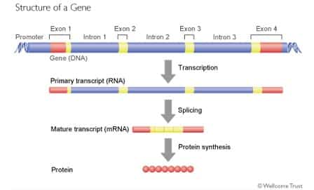
It is well-known that many eukaryotic pre-mRNA molecules have both introns and exons. The introns are removed from the mRNA, the exons are connected, and then the gene is expressed. A common question has passed through the minds of many. What exactly is the point of introns? What is their function? This article helps to answer these questions.
Introns have been called "junk DNA" but the fact that they are so prevalent and and have been preserved during evolution leads researchers to believe that introns serve some function.
 Ashok Bidwai, an assistant professor in the department of biology at West Virginia University, explained how it is widely believed that introns are remnants of genetic sequences that once served as spacers between the stretches of DNA that coded for specific, simpler proteins. As complex proteins were being evolved, regions of the genetic code (domains) may have been shuffled and brought together to generate new sequences that code for protein structures that took on new functions. This hypothesis is based on the observation that the relative positions of introns in genes remain largely the same in organisms from Drosophila melanogaster (the fruit fly) to Caenorhabditis elegans(a widely studied nematode) to mice and humans.
Some researchers have proposed that introns serve as a mechanism that selects for genes that will be expressed early during the development of an organism. There has not been much experimentation to support this hypothesis so its plausibility is uncertain.
Sandro J. de Souza, who works in Walter Gilbert's laboratory at Harvard University, expanded on the prevailing intron hypothesis. There are at least five different types of introns. Some are ribozymes. Some of these ribozymes can splice themselves out of the original transcript. The most common type of intron is called a spliceosomal or nuclear intron.This type of intron is the one found in the nuclear genes of humans.
Generally, nuclear introns are ubiquitous in complex eukaryotes. However, simple prokaryotes and eukaryotes lack introns. In complex multicellular organisms, introns are about ten times longer than the exons. The sequence and length of introns vary rapidly over evolutionary time.
Introns do sometimes have identifiable functions. Scientists have found examples of "functional nuclear introns" that can accommodate sequences important for the expression of the gene on which the intron resides. This function is not a common feature of introns, though. There are also cases in which introns contain genes for small nuclear RNA. Nuclear introns can also be important in alternative splicing, which produces multiple types of messenger RNA from a single gene. Although these examples demonstrate roles for introns, they do not explain why introns are so ubiquitous in our genes.
Introns have been called "junk DNA" but the fact that they are so prevalent and and have been preserved during evolution leads researchers to believe that introns serve some function.
Some researchers have proposed that introns serve as a mechanism that selects for genes that will be expressed early during the development of an organism. There has not been much experimentation to support this hypothesis so its plausibility is uncertain.
Sandro J. de Souza, who works in Walter Gilbert's laboratory at Harvard University, expanded on the prevailing intron hypothesis. There are at least five different types of introns. Some are ribozymes. Some of these ribozymes can splice themselves out of the original transcript. The most common type of intron is called a spliceosomal or nuclear intron.This type of intron is the one found in the nuclear genes of humans.
Generally, nuclear introns are ubiquitous in complex eukaryotes. However, simple prokaryotes and eukaryotes lack introns. In complex multicellular organisms, introns are about ten times longer than the exons. The sequence and length of introns vary rapidly over evolutionary time.
Introns do sometimes have identifiable functions. Scientists have found examples of "functional nuclear introns" that can accommodate sequences important for the expression of the gene on which the intron resides. This function is not a common feature of introns, though. There are also cases in which introns contain genes for small nuclear RNA. Nuclear introns can also be important in alternative splicing, which produces multiple types of messenger RNA from a single gene. Although these examples demonstrate roles for introns, they do not explain why introns are so ubiquitous in our genes.
Bacterial Prolyl-tRNA Synthetase
So this article talks about a specific feature of bacterial prolyl-tRNA synthetase. The article explains how the substrate specificity of bacterial prolyl-tRNA synthetase's editing domain is controlled by a tunable hydrophobic pocket. Aminoacyl-tRNA synthetases are catalysts that covalently attach amino acids onto their respective tRNA's. High accuracy in this reaction is essential to the proper decoding of genetic information for if a charged tRNA molecule contains the wrong amino acid, the incorrect amino acid will be added to the polypeptide chain, thus making a faulty protein. One way the cell tries to prevent this mishap is by proofreading newly synthesized aminoacyl-tRNA molecules. Prolyl-tRNA synthetase (ProRS) mischarges tRNA(Pro) with alanine or cysteine because of their smaller or similar sizes relative to cognate proline. Mischarged Ala-tRNA(Pro) is hydrolyzed by the editing domain, INS, present in most bacterial ProRSs. On the other hand, the INS domain is unable to deacylate Cys-tRNA(Pro). Cys-tRNA(Pro) is hydrolyzed exclusively by a free-standing trans editing domain known as YbaK.

Researchers used computational and experimental approaches to probe the molecular basis of INS domain alanine specificity. In Escherichia coli ProRS, the methyl side chain of alanine binds in a well-defined hydrophobic pocket characterized by conserved residues I263, L266, and K279 and partially conserved residue T277. Site-specific mutation of these residues leads to a significant loss in Ala-tRNA(Pro) hydrolysis, and altering the size of the pocket modulates the substrate specificity. Surprisingly, one ProRS INS domain variant displayed a complete switch in substrate specificity from alanine to cysteine.

Thursday, December 1, 2011
Chapter 11 Helpful Hints!
This video helps to conceptualize what is happening to the leading and lagging strands during transcription. The video starts out with a still picture that shows all of the relevant proteins. It then goes into an animation and shows how the leading strand is continuously synthesized, while the lagging strand is synthesized in pieces. It also shows how the lagging strand folds over so the DNA Polymerase can add nucleotides to the template strand.
This video is pretty cool! It is sooo fetch! It shows some of the processes that DNA goes through like coiling, replication, transcription and translation. It uses computer animation based on molecular research to show a 3-D view of what is happening during these amazing processes that DNA undergoes. Not only does it show the processes, but it also gives explanations about what is happening.
This video is pretty cool! It is sooo fetch! It shows some of the processes that DNA goes through like coiling, replication, transcription and translation. It uses computer animation based on molecular research to show a 3-D view of what is happening during these amazing processes that DNA undergoes. Not only does it show the processes, but it also gives explanations about what is happening.
Tuesday, November 29, 2011
The Immortal Cells from Henrietta Lacks
In order to learn and discover more about cells, researchers and even students use laboratory-grown human cells to run tests and experiments. Unfortunately for researchers, keeping a culture/cell line of human cells alive to be used over and over again was proving to be a difficult feat. After a while, the cells would just stop dividing and die off. This was the story until scientists came across Henrietta Lacks' cells. As this article explains, Henrietta Lacks' cells have proven to be "immortal".
 Henrietta Lacks was a black tobacco farmer from southern Virginia who developed cervical cancer at thirty years old. Without Henrietta's knowledge, a doctor from Johns Hopkins took a piece of her tumor and sent it to some scientists. These scientists had been trying to grow tissues in culture for decades but had had no success. They struck success with Henrietta Lacks' cells because for some reason, her cells never died.
Henrietta Lacks' cells, which have been named HeLa cells were the first humans cells to ever be grown in culture. Some of her cells achievements include playing an essential part in developing the polio vaccine and going to space in the first space mission to see what would happen to cells in zero gravity. HeLa cells have also been involved in cloning, in vitro fertilization, and gene mapping.
Over the years, there has been much confusion over the source of HeLa cells. Back in the 1950's, doctors were not too concerned with anonymity in donating samples are they are today. Since the cells are codenamed HeLa cells, when the media would get too close to finding Henrietta Lacks' family, the researcher who had grown the cells gave out the pseudonym Helen Lane, and eventually even Helen Larsen. Henrietta Lacks was not accredited for the HeLa cells until the 1970's.
Henrietta's family did not find out about the HeLa cells until twenty-five years after she died. This came about because a scientists discovered other tissue types, including prostate and breast cells, were HeLa cells. They then found out that HeLa cells could float on dist particles in the air, travel on unwashed hands, and contaminate other cultures. This created a huge controversy, so some scientists decided to track down Henrietta's family to see if they could take her family's DNA to map out Henietta's genes and found out which HeLa cell cultures belonged to Henrietta. This would help them begin to fix the problem of contamination. Since her husband only had a third-grade education he did not understand what a cell was or how scientists still had the cells from his dead wife. The scientists did not know that the Henrietta Lacks' husband and family did not understand what was going on. As a result, her family got sucked into a world of research with the cells basically taking over their lives.
Her daughter Deborah was an infant when Henrietta died. When she found out that part of her mother was still alive, she was desperate to fully understand what that meant. However, Deborah's brothers only became interested once they found out that money was involved. When they found out that their mother's cells helped launch a multi-billion dollar industry, were being sold in vials to people, and that the family got no money from it, they were furious. Most of her Henrietta's family was poverty-stricken. Therefore, her family launched a campaign to try and get what they felt they were financially owed. This campaign somewhat consumed their lives.
Bulky DNA Adducts
As this article explains, Aristolochic acids I and II are two plant toxicants found in the Aristolochiaceae plant family. The acids poison humans by messing with DNA. Metabolic activation of the aristolochic acids leads to the formation of a cyclic N-hydroxylactam product. This product can react with the peripheral amino group of purine bases, thus generating bulky DNA adducts. Therefore, these acids are classified as mutagens and human carcinogens.
 Although the AL-dG adducts would with time disappear from the DNA of laboratory animals, AL-dA lesions would still be present in the genomes of the animals. The researchers' data establish a locally perturbed double helical structure that actually accommodated the bulky adduct. The structure did this by displacing the counter residue from the bulky adducts into the major groove and stacking the ALII moiety between flanking bases. The presence of the ALII-dA perturbs the conformation of the 5'-side flanking base pair.However, all other pairs of the duplex take on their standard conformations. Thermodynamic studies have shown that the lesion slightly decreases the energy of duplex formation in a sequence-dependent manner.

Tuesday, November 15, 2011
Chapter 9 Helpful Hints!
I found this video to be quite helpful. The video explains the process of G-Protein Coupled Proteins Signal Transduction Reactions. The video takes you step by step through the process. It clearly names, labels, and identifies all of the components of the reaction and explains what is happened at each stage.
When I first clicked on this video I thought it would be a failure attempt at copying a KhanAcademy video. However, once I watched it I realized this was far from a failure. The lecture is easy to follow with many examples and labels. He takes you through the process of signal transduction in terms easy to understand, while still using all of the proper biology terminology. Many of his examples and relations are modern, easy to understand ones that further help you to comprehend the material.
When I first clicked on this video I thought it would be a failure attempt at copying a KhanAcademy video. However, once I watched it I realized this was far from a failure. The lecture is easy to follow with many examples and labels. He takes you through the process of signal transduction in terms easy to understand, while still using all of the proper biology terminology. Many of his examples and relations are modern, easy to understand ones that further help you to comprehend the material.
Apoptosis Occurs Because...
Why does apoptosis occur? Apoptosis is defined as programmed cell death, but why would cells program their own death? Also, does apoptosis occur only in higher level organisms or does it occur in organisms like bacteria and fungi as well? Luckily for us, this article answers all of these questions.
Let's attack this question by question. So, why does apoptosis occur? There are several reasons for programmed cell death. To name a few:
1. Some cells are generated in excess and only the ones that become properly functional survive. i.e. Nervous System
2. The mechanism that generates a certain type of cell luckily generates unneeded along with needed cells. Some cells that are needed die with time, but since there were extra unneeded cells produced, the individual is okay. i.e. Immune System
In essence, cells are programmed to die because they are harmful or because it takes less energy to kill them than to maintain them. Programmed cell death occurs to get rid of cells that are not needed, in the way, or potentially dangerous, as Michael Hengartner, the Senior Staff Investigator at Cold Spring Harbor Laboratory, put it.
 Now on to another question. Does apoptosis occur amongst unicellular organisms? In a unicellular organism apoptosis could be akin to suicide! Nonetheless, studies have shown processes that scientists consider to be apoptosis in single-celled organisms. The death of the mother cell during sporulation (the process of creating spores) could be considered to be programmed cell death. Some parasites, like trypanosomes change form to escape the immune response from their host. The organisms that fail to change shape just kind of die off.
Let's attack this question by question. So, why does apoptosis occur? There are several reasons for programmed cell death. To name a few:
1. Some cells are generated in excess and only the ones that become properly functional survive. i.e. Nervous System
2. The mechanism that generates a certain type of cell luckily generates unneeded along with needed cells. Some cells that are needed die with time, but since there were extra unneeded cells produced, the individual is okay. i.e. Immune System
In essence, cells are programmed to die because they are harmful or because it takes less energy to kill them than to maintain them. Programmed cell death occurs to get rid of cells that are not needed, in the way, or potentially dangerous, as Michael Hengartner, the Senior Staff Investigator at Cold Spring Harbor Laboratory, put it.

G-Protein Coupled Receptors and Cardiovascular Therapies
This article explains how G-protein coupled receptors are involved in many types of cardiovascular therapies. Adenosine, a purine nucleoside, activates four G-protein coupled receptors. There four receptors are A1, A2a, A2b, and A3. Activation of myocardial A1 receptors have been shown to inhibit a variety of myocardial pathologies associated with ischemia and reperfusion injury, These pathologies include stunning, arrhythmogenesis, coronary and ventricular dysfunction, acute myocardial infarction, apoptosis, and chronic heart failures. These findings imply several options for new cardiovascular therapies for diseases, like angina pectoris, control of cardiac rhythm, ischemic injury during an acute coronary syndrome, and heart failure.
 The main problem arising involving using full A1 agonists for these problems is the wide range of side effects. This is because of the broad physiologic spectrum of cardiac and extracardiac effects caused by the A1 receptor. This can be overcome by using partial A1 agonists. Partial A1 agonists can be used to trigger only some of the physiological responses of receptor activation. The responses triggered depend on the endogenous adenosine levels and on receptor reserves in different tissues.

Thursday, November 3, 2011
Chapter 7 Helpful Hints!
Picture this dearies. It's freshman year and we're about to have a test on cellular respiration. Now cellular respiration was very scary to me since it was complex and what have you. So, in my freshman youth, I searched "cellular respiration song" on Youtube. Alas, the first video I stumbled upon was this video. Now if you sat next to me on the day of the test you might recall me softly humming this. Fast forward two years later and I still remember this song/rap because it was so helpful! The people in the video may be... different, but they nonetheless helped me to remember a lot of the information on cellular respiration. So... enjoy!
Here's another helpful hint! So this song/rap is surprisingly very well done! They go into details that many songs leave out and use many terms that other videos leave out as well. They also go into lipolysis which we briefly touched on, which is when fatty acids are used rather than glucose molecules. So enjoy and brush up on some terms!
Here's another helpful hint! So this song/rap is surprisingly very well done! They go into details that many songs leave out and use many terms that other videos leave out as well. They also go into lipolysis which we briefly touched on, which is when fatty acids are used rather than glucose molecules. So enjoy and brush up on some terms!
Post-Mortum Respiration Leads to Autophagy Gone Wrong?
When a person dies, the person obviously is not able to and therefore, no longer breathing. However, what about the person's cells? Although, the person is no longer respiring, are the person's cells still respiring? Luckily for us, this article answers that question. As of now, it is believed that cell metabolism likely continues for four to ten minutes after a person dies, depending on the temperature around the body. Blood stops circulating, so cellular respiration can only continue for a little while. The oxygen present is used in cellular respiration, and the waste product carbon dioxide is created.

Carbon dioxide becomes carbonic acid, thus lowering the pH of the cell. The acidic environment results in the rupture of intracellular membranes. This rupturing includes the membranes of the lysosomes. Lysosomes contain enzymes which digest all sorts of macromolecules, like proteins, fats, and nucleic acids. Once the lysosome membranes are burst the cell literally begins eating itself from the inside out, resulting in the death of the cell. This process is known as autolysis.

Carbon dioxide becomes carbonic acid, thus lowering the pH of the cell. The acidic environment results in the rupture of intracellular membranes. This rupturing includes the membranes of the lysosomes. Lysosomes contain enzymes which digest all sorts of macromolecules, like proteins, fats, and nucleic acids. Once the lysosome membranes are burst the cell literally begins eating itself from the inside out, resulting in the death of the cell. This process is known as autolysis.
Wednesday, November 2, 2011
Plants' Homeostatic Reponse to Hypoxia
Plants and animals are aerobes, and therefore require in order to to respire and for energy production. This article explains how the plant response to hypoxia (decline in oxygen availability) is a bit different from the animal response to hypoxia. The decline in oxygen triggers a change in gene transcription and messenger RNA that promote anaerobic metabolism, ergo sustaining substrate-level ATP production. Furthermore, oxygen sensing has not been ascribed to a mechanism of gene regulation in response to oxygen deprivation, like it is in animals. In studying Arabidopsis, researchers showed that the N-end rule pathway of targeted proteolysis acted as a homeostatic sensor of severe low oxygen levels in the plant, through its regulation of key hypoxia-response transcription factors. Researches also found that plants lacking components of the N-end rule pathway expressed core hypoxia-response genes and were more tolerant of hypoxic stress. Hypoxia-associated ethylene response factor group VII transcription factors of Arabidopsis were identified as substrates of this pathway. Enhanced stability of one of the proteins, HRE2, under low oxygen conditions improved hypoxia survival. It also revealed a molecular mechanism for oxygen sensing in plants by the evolutionarily conserved N-end rule pathway.


Thursday, October 20, 2011
Newest Biofuel: Cow Enzymes?
Since we've realized that the waste from our excessive oil usage is detrimental to the environment, and that our oil reserves are quickly fading, scientists have been trying to think of ways to use more biofuels. Who would have thought that as this article explains, we would be looking inside of a cow's stomach?
 One of the main focuses in making biofuels is cellulosic plant matter. The break down of the large molecule, cellulose, would provide the energy that we would then harness and use for whatever needs we have. Making biofuel from cellulosic plant matter would be more environmentally friendly and economical because many cheap plants contain lots and lots of cellulose (i.e. switchgrass, Miscanthus, woodchips). The only problem with this method is that the methods for breaking down cellulose are extremely expensive.
Engineers have already been using enzymes from animals like termites to degrade through tough plant material. Unfortunately, the enzymes currently available efficient enough to make the cellulose-to-fuel conversion worthwhile. The director of the U.S. Department of Energy's Joint Genome Institute, Eddy Rubin,and sixteen colleagues discovered nearly 30,000 new enzyme candidates by analyzing DNA collected from a cow's rumen, which is first compartment in the animal's four-section stomach. The researchers were able to identify 27,755 genes that were good enough to be candidates toward cellulosic biofuel practices. They then chose 90 of the candidate genes, expressed them to produce the enzymes they code for, and then applies the enzymes to the biofeul feedstocks Miscanthus and switchgrass. Over half of the selected 90 showed the capacity to degrade at least one of the feedstocks. Researchers feel that this suggests the original, larger pool of candidates is "highly enriched" with enzymes whose activity could be useful in biofuel production.

Engineers have already been using enzymes from animals like termites to degrade through tough plant material. Unfortunately, the enzymes currently available efficient enough to make the cellulose-to-fuel conversion worthwhile. The director of the U.S. Department of Energy's Joint Genome Institute, Eddy Rubin,and sixteen colleagues discovered nearly 30,000 new enzyme candidates by analyzing DNA collected from a cow's rumen, which is first compartment in the animal's four-section stomach. The researchers were able to identify 27,755 genes that were good enough to be candidates toward cellulosic biofuel practices. They then chose 90 of the candidate genes, expressed them to produce the enzymes they code for, and then applies the enzymes to the biofeul feedstocks Miscanthus and switchgrass. Over half of the selected 90 showed the capacity to degrade at least one of the feedstocks. Researchers feel that this suggests the original, larger pool of candidates is "highly enriched" with enzymes whose activity could be useful in biofuel production.
Helpful Hint: Oxidation and Reduction...
When looking at simple problems, redox reactions make perfect sense to me. As we all know, the body and life are not simple processes therefore most reactions are not quite simple, so the more complex problems have confused me a bit. I personally always love to watch Khan Academy videos in any subject applicable because they are just so helpful! So when I found this video I was ecstatic! This video has helped me a great deal because it really breaks down what is happening in redox reactions and takes you step by step when explaining what occurs. The video also gives little hints on ways to remember certain terms and concepts. The video basically is a lecture, but it is in no way boring. I also like this video because it incorporates concepts we have already learned and mastered this year (i.e. electronegativity) to explain the oxidation and reduction reactions. So, this video will help you understand the concepts more if you're a bit confused. It will also help if you want to watch a quick lecture to refresh or confirm the concepts in your mind!
Fatty Acid Oxidation Prevents Obesity in Ob/ob Mice!!
So this article talks about how scientists have found that when endogenous ligands sustain the activation of PPAR{alpha}, hepatic fatty acid oxidation is increased, resulting in the prevention of obesity in ob/ob mice. An ob/ob (or obese) mouse is a mutant mouse that eats excessively and becomes extremely obese.
 Mice that lacked acyl-CoA oxidase 1 (Acox1), the first enzyme of the peroxisomal fatty acid β-oxidation system, are characterized as having an increased energy expenditure and a lean body phenotype. This is because of sustained activation of peroxisome proliferator-activated receptor α (PPARα) by endogenous ligands in liver that remain unmetabolized in the absence of Acox1.Scientists wanted to see what would happen if an ob/ob mice lacked Acox1. So, they made generated some ob/ob mice deficient in Acox1 and conducted tests comparing them to regular ob/ob mice.
When compared, the regular ob/ob mice were severely obese and had more epididymal fat content. The reason why the Acox1 deficient ob/ob mice were more resistant to obesity is because there is an increase in PPARα-mediated up-regulation of genes involving fatty acid oxidation in liver. The activation of PPARα in Acox1-deficient ob/ob mice also reduced the serum glucose and insulin levels, and improved glucose tolerance and insulin sensitivity. There are negatives as well though. For example, Acox1 deficient ob/ob mice manifested hepatic endoplasmic reticulum stress and also develop hepatocellular carcinomas.

When compared, the regular ob/ob mice were severely obese and had more epididymal fat content. The reason why the Acox1 deficient ob/ob mice were more resistant to obesity is because there is an increase in PPARα-mediated up-regulation of genes involving fatty acid oxidation in liver. The activation of PPARα in Acox1-deficient ob/ob mice also reduced the serum glucose and insulin levels, and improved glucose tolerance and insulin sensitivity. There are negatives as well though. For example, Acox1 deficient ob/ob mice manifested hepatic endoplasmic reticulum stress and also develop hepatocellular carcinomas.
Friday, October 7, 2011
Wee for a Wii and Quirky Quasicrystals
The Sacromento radio station KDND 107.9 held a contest in early 2007 called "Hold Your Wee for a Wii". The person who could drink the most amount of water without using the bathroom would win the Wii. Jennifer Strange, a healthy 28-year old, participated in this contest, and unfortunately lost her life because of it. Jennifer Strange died of water intoxication just hours after the contest.
Water intoxication, as explained by this article, causes problems in electrolyte balance, resulting in a rapid decrease in serum sodium concentration and eventually leads to death. The decrease in serum sodium concentration is known as hyponatremia. As we learned in class, our cells prefer isotonic solutions. With the intake of too much water, Strange's extracellular fluid became more diluted, causing her cells to be bathed in a hypotonic solution. Water flowed into her cells to try and even out the concentration of solute to solvent. This caused her cells to swell. On the way home from the radio station, Strange was complaining of a terrible headache. This is because her brain cells contained too much water, causing intracranial pressure to increase and her to experience a headache.
This article continues to explain more symptoms of water intoxication. The electrolyte imbalance previously described also results in an irregular heartbeat, can allow fluid to enter the lungs, and not only puts pressure on brain cells, but also nerve cells. All of the symptoms are caused by the swelling of the cells. In theory, the excessive amount of water in the cell could cause it to burst.
If one is beginning to experience water intoxication, one way to quickly get it under control is to take diuretics so that one can urinate. Water intoxication should be treated promptly because it leads to damage in basically every part of the body, strokes, comas, and death.
*INSERT FETCH TRANSITION HERE*
Now, let's move on to quasicrystals! As this article explains, up until 1982 scientists believed they knew when a crystal was not a crystal. Then in 1982, came this Israeli physicist named Daniel Shechtman. He discovered these things called quasicrystals in metals. These quasicrystals were crystals with odd structures that scientists had thought were impossible structures for crystals to have. At first, his discovery of quasicrystals resulted in Schectman losing his job and drawing scorn from several colleagues. Now, his discovery has earned him the 2011 Nobel Peace Prize in Chemsitry.
So what exactly are quasicrystals? First, it would help to know what a regular crystal was. Crystals are a form of matter that in which atoms are arranged in orderly patterns that repeat themselves. In order to be a crystal, the arrangement of atoms must be symmetrical when viewed from different angles. For example, each atom could be in the middle of a triangle formed by its neighbors. When turned, the series of overlapping triangles form a pattern that reappears with every 120 degrees of rotation. Atoms centered in a pattern of rectangles, squares, hexagons also exhibit this kind of symmetry, but at different angles. However, atoms in patterns made up of geometric shapes with five or more than seven sides, don't show symmetry at any angle. Ergo, materials with those arrangements were not considered crystals
So one day Schectman was mixing molten aluminum and manganese and cooling it quickly to study the properties of the resulting alloy. When he viewed the sample through a microscope he was flabbergasted! The atoms were arranged in pentagons. These pentagons were arranged in concentric circles each having ten dots. The geometric shape formed by the ten dots was a repeated pattern at 36 degrees but the pentagons were not. Schectman had just stumbled upon quasicrystals! At first, even Schectman did not believe his results. However, he realized the results were very real and after finding three highly respected coauthors, he published a paper about quasicrystals in 1984.
Today quasicrystals are still being studied but have been already put into use because of their unique properties. For example, researchers have been trying incorporate their unique properties into technologies as advanced as light-emitting diodes and as common as non-stick frying pans.
The figure on the left would be a crystal arrangement, the figure on the right would be a quasicrystal arrangemnt.

Wednesday, October 5, 2011
Great Googly Mogly, It's Glycosylation!
This article explains the process of glycosylation. Glycosylation is the process by carbohydrates are attached to protein molecules. It is a common post translational modification for protein molecules that are a part of the plasma membrane. Glycosylation involves the linking of monosaccharide units to amino acid chains. This sets up the stage for a series of enzymatic reactions which results in the formation of glycoproteins. There are 16 known enzymes that facilitate these enzymatic reactions. A typical glycoprotein has at least 41 bonds involving 8 amino acids and 13 different monosaccharide units. The major sites of protein glycosylation are the ER, Golgi , nucleus and cytoplasm.
There are 2 main types of gylcosylation.
 N-Linked Glycosylation- N-Linked Glycosylation begins with the addition of a 14-sugar precursor to an asparagine amino acid. It contains glucose, mannose and n-acetylglucosamine molecules. The whole entity is then transferred to the ER lumen. Oligosaccharyl transferase enzyme attaches the oligosaccharide chain to asparagine in the tripeptide sequence, Asn-X-Ser or Asn-X-Thr where X can be any amino acid other than Proline. The oligosaccharide attached protein sequence will now fold correctly and is translocated to the Golgi. Mannose residue is removed in the golgi apparatus.
 O-Linked Glycosylation- O-linked glycosylation begins with the addition of N-acetyl-galactosamine to the molecule by an enzyme and other carbohydrates to serine or threonine residues. O-linked glycosylation occurs at a later stage in protein processing, probably in the Golgi apparatus.
There are 2 main types of gylcosylation.


Wednesday, September 28, 2011
Chapter 4 Helpful Hints/ Human Plasma Proteome
Chapter 4: Helpful Hints
This video is a song/rap about cells and their organelles. The video is set to the song "Empire State of Mind" by Jay Z, so the song goes to a popular tune that is easy to remember. There are lots of diagrams, pictures, and labels so one can identify the parts while hearing/singing about them. Lastly, the video shows both eukaryotic and prokaryotic cells, so one can identify the cell types and structures in both types.
So this video is about endosymbiosis. Endosymbiosis is how it is believed that mitochondria and chloroplasts ended up in cells. This video has clay figures clearly labeled to represent the large and small cells involved. It also has words describing what is happening during endosymbiosis. Last but certainly not least, it has "You've Got a Friend in Me" playing in the background which relates to the video and is also such a great song!
Human Plasma Proteome
This article talks about how the human plasma proteome holds a world of answers involving disease diagnosis and therapeutic monitoring. The plasma proteome is not the only proteome being studied, but it has the largest and deepest version of the human proteome. The plasma proteome not only included plasma proteins, but also included tissue proteins and multiple distinct immunoglobin sequences.
 Before, standard proteomic technology only allowed for scientists to study 289 plasma proteins. However, new technologies are guaranteeing twice the amount of proteins to be studies in the near future. Research suggests that among these numerous plasma proteins are indicators of many human diseases. However, very few proteins are used in routine clinical diagnosis, and the rate of introduction of new protein test approved by the FDA has declined in the past ten years to one new protein per year.
This video is a song/rap about cells and their organelles. The video is set to the song "Empire State of Mind" by Jay Z, so the song goes to a popular tune that is easy to remember. There are lots of diagrams, pictures, and labels so one can identify the parts while hearing/singing about them. Lastly, the video shows both eukaryotic and prokaryotic cells, so one can identify the cell types and structures in both types.
So this video is about endosymbiosis. Endosymbiosis is how it is believed that mitochondria and chloroplasts ended up in cells. This video has clay figures clearly labeled to represent the large and small cells involved. It also has words describing what is happening during endosymbiosis. Last but certainly not least, it has "You've Got a Friend in Me" playing in the background which relates to the video and is also such a great song!
Human Plasma Proteome
This article talks about how the human plasma proteome holds a world of answers involving disease diagnosis and therapeutic monitoring. The plasma proteome is not the only proteome being studied, but it has the largest and deepest version of the human proteome. The plasma proteome not only included plasma proteins, but also included tissue proteins and multiple distinct immunoglobin sequences.

Monday, September 26, 2011
Proteins and Huntington's Disease
This article talks about the importance of protein folding and a few of the diseases that occur because of improper protein folding or a cell's protein quality control not being as strong.. Protein synthesis is essential for life, but it's not the only step. In order for proteins to function properly, they need to fold in the correct manner. One simple error in folding can lead to a completely inactive protein.
An example of this would be Huntington's Disease. Huntington's Disease (HD) is a dominantly inherited autosomal neurodegenerative disorder. It is characterized by the progressive development of mood disturbances, behavioral changes, involuntary choreiform movements, and cognitive impairments. Onset usually occurs in adulthood, and HD usually lasts 15-20 years before ending a person's life in a premature death.
What cause HD is the expansion of an unstable CAG repeat which encode glutamines close to the 5'-end of the gene for the huntingtin protein. The length of the CAG determines the phenotype of HD. The protein huntingin is found predominantly in the cytoplasm. It's function is not yet fully understood yet, but may involve cytoskeletal function or vesicle recycling. It has also been proposed that the gene may be transported to the nucleus and serve a role in the regulation of gene transcription, but this is not yet certain. The toxicity of huntingin may be caused by mutant full-length proteins or cleaved proteins. The Huntington gene is expressed in all cells but only affects a subset of neurons.
Saturday, September 24, 2011
Tay-Sachs Disease
Tay-Sachs disease is a terminal disease of the nervous system. It was first identified in 1881. Tay-Sachs disease occurs because of problems with lysosomes. Lysosomes are organelles in cells which essentially process waste. They are kind of like the garbage men of the cell! I came up with that analogy all by myself! Clever, I know.
This article explains how a person with Tay-Sachs has mutated genes that result in the production of enzymes less effective in breaking down gangliosides. These enzymes are found in lysosomes. Gangliosides are fatty cell products. Since these enzymes are less effective, gangliosides build up in the lysosomes and eventually overload the cell. This build up causes damage to nerve cells.
This mess up on a cellular level results in terrible outcomes for people with Tay-Sachs disease. This article talks about symptoms of the disease include deafness, blindness, decreased muscle tone, loss of motor skills, loss of muscle function/paralysis, and many others. Tay-Sachs is an inherited genetic disease. The child must receive the gene from both parents in order to be affected by it, otherwise the child will just be a carrier. This is why nerve damage caused by the disease often begins when the child is still in the womb. Symptoms usually start popping up when the child is 3 to 6 months old. Unfortunately, most people with Tay-Sachs disease don't live past 4 or 5 years old.
Tuesday, September 20, 2011
Chapter 3: Hunky-Dory Proteins and Nucleic Acids!
This blog will be based off of the virtual lectures on proteins and nucleic acids.
Proteins
Proteins are quintessential in our survival. They have so many functions in a cell, and thus in our lives. Proteins are used for support, as enzymes (speeding up biological reactions), membrane transport, and producing cellular movements. Amazingly, proteins functions don't stop there! Proteins are also regulators, receptors, hormones, antibodies, venom and toxins, and for storage. Proteins do so many things that allow us to live.
 Proteins are able to do so many things because they are built differently. The structure of a protein determines its function. There are four levels of protein structure. These four structures are the primary structure, secondary structure, tertiary structure, and quaternary structure.
Tertiary Structure: The tertiary structure is the overall three-dimensional shape of a protein. As each secondary structure takes hold, the protein takes a globular shape. In an aqueous (polar) environment, the folding into a compact, globular structure would be to "hide" the hydrophobic amino acids by putting them in the interior, and exposing the hydrophilic heads to the exterior aqueous environment. In the tertiary structure, the R-groups are what stabilize the structure. Proteins can denature, or lose their structure and function. Proteins are function when folded, so when they are denatured, they become unfolded and inactive. Denaturation can be caused by an increase in heat, change in pH levels, and/or the addition of salt.
Quaternary Structure: Quaternary structures occurs when two or more proteins join together. This forms a larger, more complex protein.
Proteins exist all throughout our cells and bodies. Their many structures and functions keep us going and alive everyday.
A nucleoside only consists of the sugar and nitrogen base. A nucleoside with one phosphate group is referred to as nucleoside monophosphate. It is referred to as a nucleoside diphosphate and nucleoside triphosphate with 2 and 3 phosphates, respectively.
There are major differences between RNA and DNA. RNA is usually found in one strand rather than in a double-helix like DNA, and has a ribose sugar rather than a deoxyribose sugar. In RNA, uracil replaces thymine, which is found in DNA. Lastly, RNA is synthesized from a DNA template.
Helpful Hints!
Article
Proteins
Proteins are quintessential in our survival. They have so many functions in a cell, and thus in our lives. Proteins are used for support, as enzymes (speeding up biological reactions), membrane transport, and producing cellular movements. Amazingly, proteins functions don't stop there! Proteins are also regulators, receptors, hormones, antibodies, venom and toxins, and for storage. Proteins do so many things that allow us to live.

Primary Structure: The primary structure of a protein is the sequence of amino acids that characterizes a specific protein. The amino acids are joined together by dehydration synthesis. The bonds are commonly referred to as peptide bonds. There are about 20 amino acids. The sequence of these amino acids in a polypeptide is what determines its function. Of these amino acids some are acidic, some are basic, some are polar, and some are non polar.
Secondary Structure: The secondary structure occurs when amino acids interact with one another. They bend/twist and form hydrogen bonds with one another. Certain secondary structure have distinctive shapes and have been named alpha-helix and beta-strands. Secondary structures are stabilized by the presence of hydrogen bonds.Tertiary Structure: The tertiary structure is the overall three-dimensional shape of a protein. As each secondary structure takes hold, the protein takes a globular shape. In an aqueous (polar) environment, the folding into a compact, globular structure would be to "hide" the hydrophobic amino acids by putting them in the interior, and exposing the hydrophilic heads to the exterior aqueous environment. In the tertiary structure, the R-groups are what stabilize the structure. Proteins can denature, or lose their structure and function. Proteins are function when folded, so when they are denatured, they become unfolded and inactive. Denaturation can be caused by an increase in heat, change in pH levels, and/or the addition of salt.
Quaternary Structure: Quaternary structures occurs when two or more proteins join together. This forms a larger, more complex protein.
Proteins exist all throughout our cells and bodies. Their many structures and functions keep us going and alive everyday.
Nucleic Acids
Nucleic acids are long polymers of nucleotide building blocks. The two types of nucleic acids are DNA (Deoxyribonucleic Acid) and RNA (Ribonucleic Acid). DNA stores our hereditary information, which includes all of the information for our cells to function properly. RNA is used in various forms to assemble proteins. Nucleotides are made up of a five-carbon sugar, a nitrogenous base, and three phosphate groups. In a DNA nucleotide, the 2' carbon only has Hydrogen (Hence the deoxy), while RNA has a hydroxyl (hydrogen and oxygen) at it's 2' carbon.A nucleoside only consists of the sugar and nitrogen base. A nucleoside with one phosphate group is referred to as nucleoside monophosphate. It is referred to as a nucleoside diphosphate and nucleoside triphosphate with 2 and 3 phosphates, respectively.
There are 4 nucleotides used to construct DNA and 4 nucleotides used to construct RNA. DNA consists of the nucleotides adenine (A), thymine (T), cytosine (C), and guanine (G). RNA consists of the nucleotides adenine (A), uracil (U), cytosine (C), and guanine (G). What varies in nucleotides is the nitrogenous base. The two categories of nucleotides are pyrimidines and purines. Pyrimidines only have one ring and purines have two rings.
The double-helical structure in DNA is caused by the formation of hydrogen bonds. Phosphodiester bonds are covalent bonds found specifically in nucleic acids. The phosphodiester bonds are what link the two, antiparallel strands together. One nucleotide is linked to the next nucleotide by dehydration synthesis. In DNA, A links with T, and C links with G. In RNA, A links with U, and C links with G. DNA has opposite ends. One end is 5' and the other is 3', based off of which sugar is on what end. As previously stated, DNA is antiparallel. This is because one strand runs in the 5' to 3' direction, while the other runs in the 3' to 5' direction. There are major differences between RNA and DNA. RNA is usually found in one strand rather than in a double-helix like DNA, and has a ribose sugar rather than a deoxyribose sugar. In RNA, uracil replaces thymine, which is found in DNA. Lastly, RNA is synthesized from a DNA template.
Helpful Hints!
Alright so this video is a song about DNA set to the tune of (You Drive Me) Crazy by Britney Spears. The only downside is you can't hear anyone singing, but the lyrics are there for you to follow. I guess it's like a karaoke thing... I wasn't going to include it as a helpful hint, but the lyrics actually fit into the song and make you memorize some DNA facts in fun, creative ways!
This video is extremely informative. The structure of DNA is explained in detail, yet is still understandable. The DNA is examined all the way down to atomic level, but it does not over-complicate the subject. If one had a few questions/uncertainties about DNA, or wanted a quick refresher, this would be a great video to watch!
Article
This article, talks about how there's increasing evidence showing that cancer cells. acquire "stem-like" epigenetic and signaling characteristics during the tumorigenic process. This includes global DNA hypo-methylation, gene-specific DNA hyper-methylation, and small RNA deregulation. In both stem cells and in differentiated cells, RNA has been shown to be an epigenentic regulator. Piwi-interacting RNAs (piRNAs), maintains genome integrity by epigenetically silencing transposons by DNA methylation. The human Piwi ortholog (a protein), Hiwi, has been found to be expressed in many human cancers. Unfortunately, there has not been much investigation about the role Piwi and piRNAs might play in contributing to the "stem-like" epigenetic state of a cancer.
Subscribe to:
Posts (Atom)